// blog
Four Linked Views of a Scan in Your Browser: Slice, Sagittal, Coronal and 3D
Our free DICOM viewer now has layouts. Image + 3D shows the slice with the 3D view beside it; Four views shows the slice, a sagittal cut, a coronal cut and 3D together. The views are linked: scroll the slices and a red outline moves through the 3D view; click a cut, the 3D view, or the image with Crosshair, and every view goes to that spot. The cuts are read from the original images at full resolution, you can measure lengths on them, and a double-click shows any view big. It runs in the browser, on your own files or a PACS study, and nothing is uploaded.

Imaging workstations can show a CT or MRI four ways at once: the slice, a cut from the side, a cut from the front, and 3D. Click a spot and all four go there.
Our free DICOM viewer does that now, in the browser, with nothing to install and nothing uploaded.
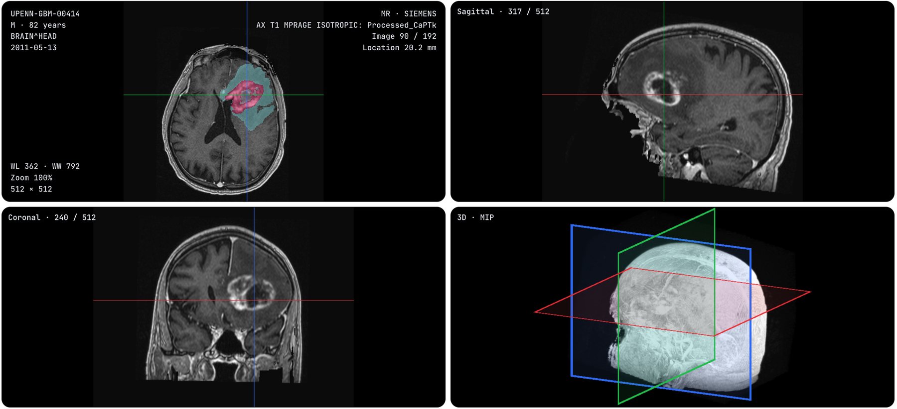
A public brain MRI and its AI segmentation, opened from the NCI Imaging Data Commons. Every line and plane in one color is the same cut in every view.
Two layouts
Open a CT or MRI series and pick from Layout:
- Image + 3D. The slice is big, the 3D view beside it. Scroll the slices and a red outline moves through the volume, so you always see where you are. Swap makes the 3D view the big one.
- Four views. The slice, a sagittal cut (from the side), a coronal cut (from the front) and 3D.
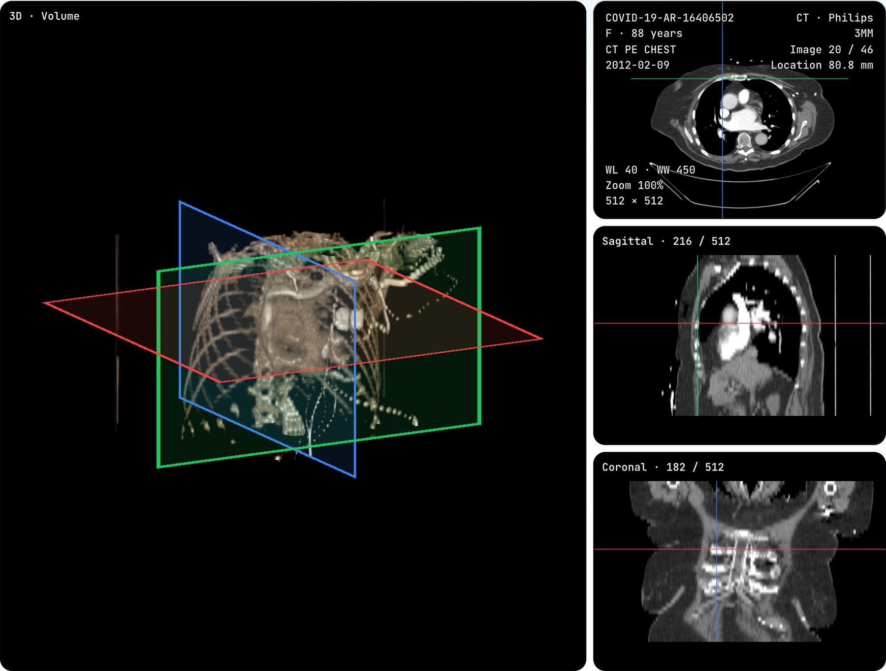
Four views with the 3D view shown big: a double-click on any view does it.
Click anywhere, everything follows
The views point at one spot, and each cut keeps its color everywhere:
- Blue is the sagittal cut, green the coronal cut, red the slice.
- Click or drag on a cut, and the other views move there.
- Click the 3D view, and the slice through what you clicked opens, with the spot marked.
- Crosshair does the same on the slice itself.
- The mouse wheel over a cut moves that cut, one image row at a time.
Made for real work
- Sharp cuts. The sagittal and coronal views are read from the original images at full resolution, 512 pixels across on a typical CT, not from a reduced 3D copy.
- Measure on the cuts. With Measure, drag on a cut for a length in millimeters. Click it and Delete removes it.
- Any view big. Double-click a view to show it big beside the other three; double-click again for four alike.
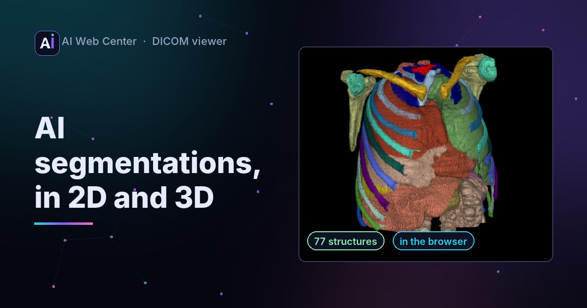
- Everything else still works: window presets, Volume, MIP and Planes in 3D, AI segmentations in color, reports and their measurements, export to PNG or JPEG.
- On a phone the views stack, the slice first.
Try it
On the sample: open the DICOM viewer, pick the Chest CT, and choose Layout, then Four views.
On the scan in the picture:
- Choose Open, then Open from a PACS, and press Try a public PACS.
- Search Patient ID
UPENN-GBM-00414, a public research MRI. - Open the MRI series. Its AI segmentation comes with it.
- Choose Layout, then Four views, and click around.
On your own scan: drop the folder from your CD or download on the viewer. It’s read in your browser and never uploaded.
Also new this week
- Reports beside the images. A measurement report from a person or an AI tool is drawn on the scan. How it works.
- Delete one measurement. Click a line, angle or ellipse, then Delete.
- An Export menu. Export to PNG, or a smaller JPEG for email.
- Subtraction (DSA) for angiography runs: pick a mask frame and the vessels filling with contrast stand out.
Private, and only a viewer
The images are read in your browser. Nothing is uploaded, and from a PACS your browser asks the archive directly. The viewer is a technology demonstration, not a medical device, and it doesn’t interpret images.
Building imaging into your own portal or product? Tell me about it.
Frequently asked questions
What are sagittal and coronal views?
Two other directions through the same stack of images. A CT or MRI is usually taken as axial slices, across the body. A sagittal cut runs from front to back, as if seen from the side; a coronal cut runs from left to right, as if seen from the front. The viewer builds both from the slices you open.
Which scans can use the layouts?
A CT or MRI series of at least 16 gray images of the same size, the same series that can be shown in 3D. A single X-ray or a moving ultrasound clip stays in one view.
How do I make the 3D view the big one?
In Image + 3D, press Swap or double-click the 3D view. In Four views, double-click any view to show it big beside the other three, and double-click it again for four alike.
Are the cuts as sharp as the slices?
Across, yes: they're read from the original images at full resolution. From top to bottom a cut is only as fine as the distance between the images, so a series with thick slices looks stepped from the side.
Can I measure on the cuts?
Yes. With Measure, drag on the sagittal or coronal view for a length in millimeters. Click a length and press Delete to remove it.
Is anything uploaded?
No. The images are read in your browser; from a PACS, your browser asks the archive directly.
Can I use it for a diagnosis?
No. The viewer is a free technology demonstration, not a medical device.