// blog
DICOM vs JPEG: Why a Screenshot of Your Scan Isn't Your Scan
A DICOM file keeps every value the scanner measured (a CT typically stores 4,096 or more levels per pixel), plus details like the pixel size in millimeters and how to display it. A JPEG or screenshot is one fixed view with at most 256 gray levels and no details: you can’t change the contrast, measure, or scroll through the other slices. Send DICOM to a doctor; use a JPEG only for a picture in a slide or a message.
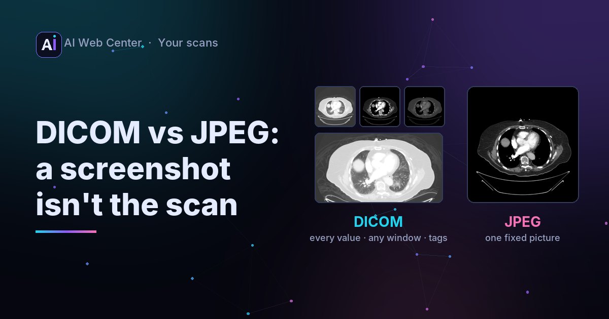
It’s tempting to share a scan the easy way: screenshot the viewer, or export a JPEG. For a picture in a message, that’s fine. For anyone who has to look at the scan properly, it throws most of it away.
What a DICOM file keeps
Take the chest CT in our viewer. Each slice is a DICOM file with:
- Every measured value. It stores 12 bits per pixel: 4,096 levels, from air to bone. Some scanners store 16.
- The pixel size: 0.9765625 mm, so distances can be measured in millimeters.
- How to display it, how to convert the values to Hounsfield units, and 103 elements in total: the scanner, the settings, the date.
- Its place in the series, so the viewer can stack the slices in order.
What a JPEG keeps
- 256 gray levels. A screen shows about that many, so the viewer has to choose which part of the 4,096 to show. That choice is the window, and the JPEG freezes one of them.
- No details. No pixel size, so no measuring. No series, so no scrolling.
- Often lossy compression, which blurs fine edges.
The picture at the top shows it: from one DICOM slice we can draw the lungs, the soft tissue or the bones. The JPEG is one of those, forever. Window and level explains how that choice works.
When a JPEG is fine
- A picture for a slide, a report you’re writing, or a message to family.
- A thumbnail in a portal or an app.
For those, convert with the right window, for example with DCMTK: dcm2img +oj +Ww 40 400 scan.dcm scan.jpg. See how to open a .dcm file.
When it has to be DICOM
- A second opinion or a new doctor. They’ll want to scroll through every slice and set their own window.
- Comparing with a later scan. Measurements need the pixel size.
- Anything going into another hospital’s system. Those import DICOM, not pictures.
Key takeaways
- DICOM keeps every value, the pixel size and every slice; a JPEG keeps one view.
- A screen can’t show all of a CT’s gray levels at once, which is why viewers have a window.
- Share JPEGs as pictures; send DICOM to doctors.
- Check a screenshot for burned-in names or dates before sharing it.
See one slice in different windows →
Run a practice or an imaging center? We’re glad to share what we know. When you want a portal that makes all of this easy for your patients, we’ll build it for you, and because we work with AI-assisted development, it costs far less than you might expect. Let’s talk.
Frequently asked questions
Can I send my doctor a JPEG of my scan?
For a second look, send the DICOM files. A doctor reading a scan changes the contrast, measures and scrolls through every slice, and a JPEG allows none of that.
Can a DICOM file contain a JPEG?
Yes. DICOM can store its pixels JPEG-compressed, and it still keeps all the details about the scan. That's different from exporting a single JPEG picture, which drops them.
Is a JPEG safer to share because it has no name in it?
A JPEG has no DICOM tags, but text burned into the image (common on ultrasound and on screenshots of viewers) can still show your name or dates. Look at the whole picture before sharing it.