Free tool · Medical imaging
DICOM viewer
Open your own DICOM files, a CD’s folder or a ZIP, a series from a PACS over DICOMweb, or pick a sample: a chest CT with an AI segmentation of 77 structures, a chest CT, a beating heart, breathing lungs, a brain MRI, a liver ultrasound clip and two chest X-rays. Work with them the way a radiology workstation does: scroll and play, drag to set the window and level, zoom, pan, invert, measure lengths and angles, and draw an ellipse for an area’s size and average value, full screen if you like, with the patient, study and acquisition details beside the image and every DICOM tag below it. Press 3D to stack a CT or MRI series into a volume you can turn and cut into. Your files are read in your browser and never uploaded, and a PACS is asked by your browser directly. The samples are public research data, prepared with the OFFIS DICOM Toolkit (DCMTK).
Your files stay on your device.
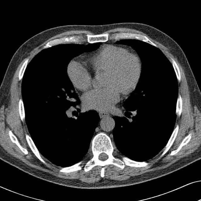
Drag: left/right changes contrast (width), up/down brightness (level). The mouse wheel or arrow keys move through the images, space plays them; Ctrl/⌘ + wheel zooms. The right mouse button changes the window (right-click Pan, Zoom or Scroll to put that on it instead; the small mouse shows which), the middle one pans; keys 1 to 9 pick a window. F or a double-click: full screen. You can also drop DICOM files, a folder or a ZIP on the image.
Made by AI Web Center · built on DCMTK v3.7.1
Need imaging in your own portal? Viewer, archive, integration.
I build web applications for clinics, imaging centers and health-tech teams: patient and referrer portals that show studies in the browser, connections to a PACS, and the APIs between them. Tell me what you need and get a first proposal.
Put the viewer on your website
Clinics, imaging centers and teaching sites can show this viewer on their own pages, free. Only the viewer, without these samples:
<div class="aiwc-dicom" data-file="/studies/ct-chest.zip"></div>
<script src="https://aiwebcenter.com/embed/dicom-viewer.js" async></script>
Your page reads the study and hands it over in the browser; visitors can also drop their own files on it, or open a study from their PACS. Setup and options.
Frequently asked questions
What is a DICOM file?
DICOM is the standard format and network protocol for medical images. A DICOM file holds the image pixels together with tags: who the patient is, what study and series it belongs to, which machine made it, and how to display it, such as the pixel spacing and the suggested window. A CT or MRI series is usually one file per slice.
What do window and level mean?
A CT stores far more shades of gray than a screen can show, so the viewer shows only a range of them. The level (window center) picks the middle of that range and the width sets how wide it is. A narrow lung window makes the airways visible; a wide bone window shows the skeleton. Drag left and right for the width, up and down for the level.
What are Hounsfield units?
CT values are measured in Hounsfield units (HU): water is 0, air is about -1000, fat is around -100, and bone is in the hundreds or more. Point at the CT and the lower right corner shows the value under the mouse in HU.
How are the distances measured?
With the Pixel Spacing tag of the file, which says how many millimeters one pixel covers. The X-rays were scaled down for the web, and DCMTK's dcmscale rewrote their pixel spacing to match, so the measurements stay in millimeters. Angles use the same spacing, and an ellipse shows its area with the average, spread, lowest and highest value inside it (HU on a CT).
Why does the CR X-ray look inverted in the file?
It is stored as MONOCHROME1, where a higher value means darker. The viewer reads the Photometric Interpretation tag and flips it, so bone is white as usual. Press Invert to see it the other way around.
Where do the images come from, and are they real patients?
The human studies are from public research collections of The Cancer Imaging Archive (TCIA): NLST (the National Lung Screening Trial) for the segmented chest CT, whose segmentation comes from the TotalSegmentator-CT-Segmentations collection of the NCI Imaging Data Commons, COVID-19-AR for the other chest CT and the X-rays, 4D-Lung for the breathing CT, UPENN-GBM for the brain MRI and B-mode-and-CEUS-Liver for the ultrasound. The archive removes names and other identifiers before publishing: the patient ID you see is its own code. The heart is a pig's, from a published animal study. All are shared under Creative Commons Attribution licenses.
What is DCMTK, and what does it do here?
DCMTK is the open-source DICOM toolkit from OFFIS in Oldenburg, Germany: C++ libraries and command-line tools for reading, writing, converting, compressing and sending DICOM files. Here it reads every tag (dcmdump), extracts the pixels, scales the large X-rays (dcmscale) and renders the preview images (dcm2img). The browser then does the window, level, zoom and measuring itself.
Does it show segmentations (DICOM SEG)?
Yes. Open the segmentation file together with its images (a folder with both, or the files side by side), and each segment is laid over the images in its own color, with a list beside the image to show or hide each one and an opacity slider; S shows or hides them all. From a PACS, opening an image series also brings the segmentations drawn on it, and opening a segmentation series brings its images. That's how outlines from a person or an AI model, such as TotalSegmentator, come back into a viewer. Binary and fractional segmentations are read. They show in 2D and in 3D: on the three crossing planes, and in the volume, where Segments only hides the scan and leaves the structures as a 3D model you can turn and cut into. The first sample, the chest CT with an AI segmentation, shows it without any files of your own.
Can I open my own DICOM files here?
Yes. Click Open files or Open a folder, or drop files or a folder on the image. A ZIP (a downloaded study or a zipped CD) opens too: it is unzipped in your browser. They are read by your browser on your own device: nothing is uploaded or stored. Close and delete removes them from the page, and they are gone when you leave or reload it too. A folder or several files open as one series (the one with the most images); files that aren't DICOM are skipped.
Which DICOM files does it open?
Uncompressed files (implicit or explicit VR, little or big endian), deflated files, RLE lossless, baseline and 12-bit JPEG, JPEG lossless, JPEG-LS, JPEG 2000 and HTJ2K, in gray (8 to 32 bits), color or palette color, single images or multi-frame clips. Segmentations, structured reports and PDFs stored as DICOM open too. A DICOMDIR is only the index of a CD: open the image files in its folders instead.
Can it open studies from a PACS?
Yes, from any PACS that speaks DICOMweb: we tested Orthanc and dcm4chee (also secured with Keycloak), and the Google Cloud and Microsoft Azure DICOM services speak the same standard. Click Open from a PACS, enter its DICOMweb address and sign in: a user name and password, or single sign-on with Keycloak or another OpenID provider, in a small sign-in window. Search by patient, ID, accession number, date, modality, study description or referring physician, page through the results, pick a study, then a series: it downloads and opens like your own files. Your browser asks the PACS directly; nothing passes through our server, and the password stays in the tab. The PACS has to allow this website (CORS): dcm4chee does already, Orthanc needs a proxy that adds it. A PACS on your local network works too; the first time, Chrome asks whether this website may reach your local network. A PACS that speaks only classic DICOM networking (DIMSE) needs a DICOMweb gateway in front of it. Try a public PACS connects to the NCI Imaging Data Commons, public research scans.
How does the 3D view work?
Press 3D on a CT or MRI series (at least 16 images of the same size, not a moving clip). The viewer stacks the images at the distance between them that the files give and puts the volume on your graphics card (WebGL2), where every pixel of the view sends a ray through it. Volume colours what the ray passes by a look: bone, soft tissue, skin or the lungs and airways for a CT, the standard or the brightest values for an MRI. MIP shows the brightest value along each ray through the window you pick, and Planes shows three crossing cuts. Drag to turn it, scroll to cut into it from the side facing you, and press Ctrl or ⌘ while scrolling, or pinch, to zoom. Like everything else here, it runs on your own device.
Why does an MRI look layered in 3D?
Many series have slices much thicker than their pixels: the brain MRI here has 0.43 mm pixels but one image every 6 mm. The viewer blends the values between two images, but it can't know what was between them, so from the side the volume shows steps. A CT with thin slices looks much smoother.
Why do the heart and the lungs move?
They are cine studies: several images of the same place at different moments, which the viewer plays in a loop at the rate stored in the file. The breathing CT is a 4D CT, sorted into ten phases of one breath and shown here as a front view rebuilt from each phase's slices. The heart MRI keeps two moments of the heartbeat, squeezed and relaxed. The ultrasound clip is 38 frames in one file.
Can I use this to read my own scans?
No. This is a technology demonstration, not a medical device, and it is not for diagnosis. Ask the clinic or hospital that made your scan for the report from a radiologist.
Can I put this viewer on my own website?
Yes, free, with two lines of HTML: a div with the study's address and one script tag. Your page reads the file and hands it to the viewer in the visitor's browser, and visitors can drop their own files on it. It also opens studies from the visitor's PACS, like this page. The embedded viewer is only a viewer, without these samples. Setup and options: https://aiwebcenter.com/tools/dicom-viewer/for-websites/
Sources
- DICOMweb (DICOM PS3.18: Web Services). QIDO-RS to search, WADO-RS to retrieve: how the viewer talks to a PACS
- NCI Imaging Data Commons. The public PACS behind Try a public PACS: de-identified research collections
- dcm4chee-arc-light wiki: Run secured archive services on a single host. The secured dcm4chee with Keycloak that the viewer's single sign-on was tested against
- Chrome for Developers: Local Network Access. Why Chrome asks before this website reaches a PACS on your local network (from Chrome 142)
- DCMTK, the OFFIS DICOM Toolkit (official GitHub mirror). Release 3.7.1, October 15, 2026; BSD-style license (COPYRIGHT); the modules and tools used here
- COVID-19-AR (The Cancer Imaging Archive). Chest CT and chest X-rays; Creative Commons Attribution 4.0
- UPENN-GBM (The Cancer Imaging Archive). Brain MRI (axial T2 FLAIR, processed); Creative Commons Attribution 4.0
- 4D-Lung (The Cancer Imaging Archive). Breathing chest CT, ten phases of a planning 4D CT; Creative Commons Attribution 3.0
- B-mode-and-CEUS-Liver (The Cancer Imaging Archive). Liver ultrasound clip, 38 frames; Creative Commons Attribution 4.0
- Cardiac Cine MRI images and labels of Sus scrofa at 7T (Lohr et al., Zenodo, 2023). Pig heart MRI, systole and diastole; Creative Commons Attribution 4.0
- Creative Commons Attribution 3.0 Unported
- Creative Commons Attribution 4.0 International